Over the years I’ve seen a number of birds with eye problems. Some of them appeared to be infections and others injuries but I’m beginning to notice a pattern of symptoms that looks similar from bird to bird, particularly in raptors. I’ve included two examples here.
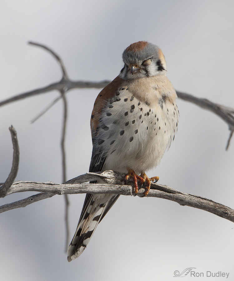
At first I thought this male American Kestrel was simply sleeping but it soon became apparent that it wasn’t opening its left eye at all. It took off after I got off a few quick shots.
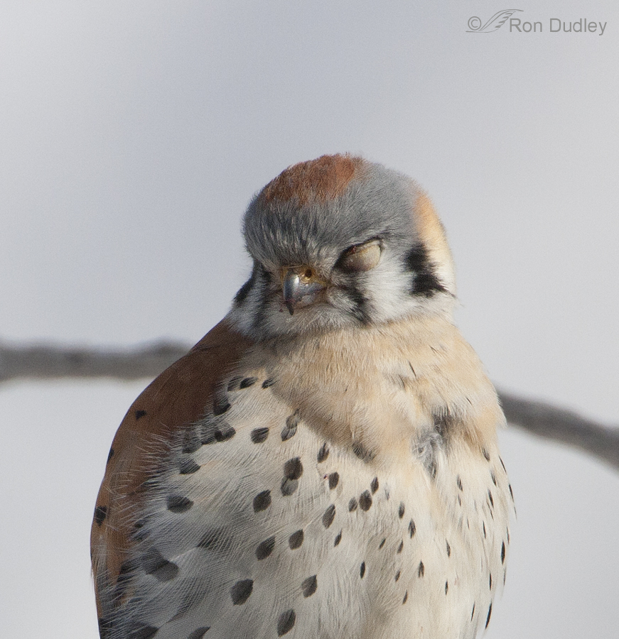
A tight crop to show more detail.
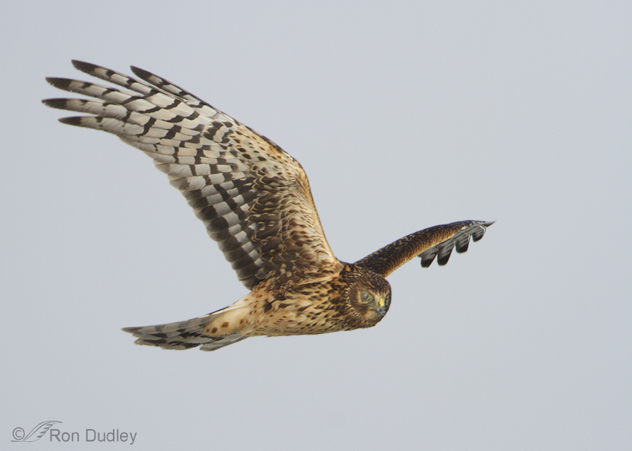
This Northern Harrier had a similar problem, this time with the right eye.
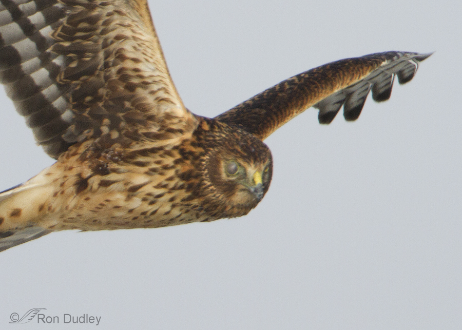
Another tight crop for more detail.
The other eye of both birds appeared normal, as did their behavior as far as I could tell. To my untrained eye it looks like the eyelids of each bird are open and the problem may have to do with either the nictitating membrane or the cornea. I’m struck by the similarity of the physical appearance of the abnormal eye in both birds and I’ve seen similar conditions in at least one other raptor (a female kestrel).
I’m always interested and curious about what I see in the field when it involves birds (good or bad) so I wonder if any of my readers have any insight as to what may be going on here. I’ve posted other images of this harrier previously and asked about the eye but I’ve just recently noticed the apparent similarities between the eye of this harrier and kestrel. Am I correct in believing that the symptoms that we can see in both birds are similar? From the physical appearance of the eyes can it be determined if these “anomalies” are likely caused by injury or infection (or perhaps injury followed by infection) or something else?
Thanks in advance for any feedback or insight.
Ron


Patty and William – there are two national/international professional associations of wildlife rehabilitators: The National Wildlife Rehabiltators Association (www.nwrawildlife.org) and the International Wildlife Rehabilitation Council (www.theiwrc.org). Neither, unfortunately, is able to collect the composite data William has suggested might be available. We’ve talked about it for years; there is one software application available, called WildONE, developed by the Wildlife Center of Virginia, that does input participants’ data into a national database, but its use is not widespread. We used it for awhile, but it’s just not as intuitive and easy to use as the one we are using now. At a conference I just attended last week in Canada, there was a talk about at least coordinating the use of certain terms for type of injury, caues, etc., no matter what software you use, so that it could eventually be consolidated, but it’s got a long way to go! COmplicated by the fact that federal reporting only covers birds but at least would be consistent – but don’t include the level of detail like ‘housefinch conjunctivitis’ as a reason for admission; state requirements vary a lot and no one really likes to do MORE than is required, paper-work wise. ANd, as Wally has said, mycoplasmal conjunctivitis (house finch conjunctivitis) started in the East and has spread not only West but to other species besides house finches – and few rehabilitators have the resources to do a definitive test of the underlying cause of conjunctivitis. We make assumptions that it might be that, but few test to be sure. Instead, we treat it as if it is.
Thanks Louise. I knew of the two organizations but haven’t had contact with them for a long time and couldn’t remember their proper names and never did know what their records might indicate or involve. Knew them for the training programs they offered to the rehabbers of N.A. Much of my learning came from the likes of Pat Redig and Kay McKeever and being a falconer.
Bill
Then you’ve learned from the best, Bill!
just reading the comments today, fascinating!
Thank you, William, for the follow-up information about the Kestrel. At the time, I wrote a note in my field journal that it appeared to be an injury directly to the eye and I speculated the coloration was due to blood in the eye. Of course, I am the furthest thing from an expert, but it seemed to fit in with Ron’s post about raptor eye injuries.
Patty, in the early 1990’s, House Finches in Maryland and Virginia were noted with mycoplasmal conjunctivitis, a respiratory ailment caused by a bacterium which affects their eyes. The eye becomes swollen, discolored and eventually crusts over. Sometimes it will clear up on its own, many times it will result in blindness and the death of the bird. This disease spread very quickly throughout the eastern U.S. and many cases are now showing up in the west
(Sorry, Ron, didn’t mean to hijack your post!)
Wally: The situation is called hyphema (blood in the anterior chamber of the eye). It often resorbs very well within a few weeks.
The eye is such a complex structure and important to a birds survival. So many things can go wrong and often a single component or issue leads to ethical considerations in the birds rehabilitation (I’m only talking raptors here but obviously often considerations of other birds as well). In my experience here are the main types of visual deficiencys:
Corneal ulceration and scarrings; hyphema; torn irisis; senechia (scarred attachments of tissue to either the lens or cornea and often result in cataracts); and detached retinas. And these are only the eye itself not the surrounding structures.
Factors of age, sex, species, presumed experience and demonstrated proficiency. This is a huge topic.
Bill
I was especially interested in Louise Shimmel’s information, as usual, and Wally’s comments about house finches. The bird we found and had treated was a house finch. I wonder if they are particularly at risk, for some reason, for eye problems. I got the idea from our vet that this was not uncommon in these birds.
Patty: Likely your vet is the best person in your life to answer your question if that is still possible. Many vet schools now offer classes in wildlife rehabilitation with some specializing in certain Classes of animals and possiblr certain genus (because of regional issues) so possibly your vet saw more house finches than say another vet might have. For other reasons as well. Population density being one.
There is an North American wide but more US active wildlife organization that many wildlife rehabbers belong to. Louise will know the organizations name. I knew of them but was never a member in the decades I have been a rehabber (only specialize in raptors). Possibly they keep records by species of contacts with rehabbers and specific types of injuries.
On the topic of how serious an eye injury can be for a raptor vs potential prey species there seems to be varying concerns for the different species abilities to survive and therefore the ethical release of various species. It is a complicated issue and the degree of injury to the eye needs to be considered. Rarely are two cases the very same.
Bill
Ron, very informative and interesting! I’ve seen quite a few House Finches suffering from conjunctivitis and recently found an American Kestrel with what I assume was an eye injury. His left eye appeared completely orange. Not sure if this may have been an injury to the pupil or if this was an eyelid closed. Based on your images, the eyelid seems as if it would be grayish. Very interesting input from your audience of experts!
My guess (dangerous to do, lol) is that the eye had some level of blood from an injury. If you have ever seen the antics that go on in a cavity or box of young Kestrels its not hard to i,agine the injuries that might occur even before fledge. Owls close their eyes by lowering the upper eyelid while the rest of the raptors close their eye by raising the lower lid (all have 2 eyelids plus a nictitating membrane). The eye lids have “lashes” at the edge but otherwise the skin is featherless. This flesh is normally pale (almost caucasian skin colored) so it is unlikely it was an eyelid you were seeing.
Bill
Thank you. And all your knowledgeable commentators.
It’s quite a group around here, isn’t it, Elephant’s Child?
Excellent information from Louise Shimmel which is much appreciated from this reader of Ron’s post.
Many thanks.
I heartily agree, Dick.
Our vet is also a licensed wildlife rehabilitatior and we took a finch to her because if an eye infection…eyes were “glued” shut ..it was staggering around “blind”…after about a week of treatment, she returned the bird to us and we released it near where we’d found it. It’s a pretty dirty world in more ways than one these days…And more possible contaminants…She told us the problem wasn’t that unusual and that “our” bird was lucky to be caught and treated.
Your were the primary component in the “luck” of that finch, Patty. Good job!
We see what we diagnose as actual conjunctivitis in very few birds – i.e., an inflammation of the conjunctiva, the membrane around and over the eye. Mostly I’ve seen it in young owls and it’s been pretty easily treatable with an ophthalmic antibiotic ointment. We also see a fair number of eye injuries, including what we suspect is trauma-induced cataracts (e.g., in nestling owls that have fallen from the nest) as well as what we suspect is age-induced cataracts, particularly in our older education birds but some birds come in with it. We’ve seen problems with the nictitans – which, if it doesn’t work to moisturize and clean the surface of the eye, leads to dry eyes and fungal or other infections. We’ve seen scratches to the cornea, punctures or lacerations … the hawk of a falconry acquaintance scratched his cornea diving into brush after a rabbit – I’m sure this happens in the wild as well and can lead to ulcerated corneas and even the loss of the eye. With these kinds of injuries, it’s not uncommon for them to keep the lid closed which, in birds, typically means the lower lid is up. Sometimes discharge from the injury leads to the crustiness that appears to show on the edge of the kestrel’s closed eye. We’ve seen barbed wire injuries to the eye and lids, including a red-tail years ago with deep pockets of inspissated pus under the eye and into the sinuses. One of our gyrfalcons was found with an eye totally covered over with pus, so badly infected it had to be removed – who knows how she did that … maybe got poked by a shorebird she was catching? Blunt trauma like collisions with windows (one of our merlins) or vehicles (many owls) can rupture the eye and cause a slow or rapid deflation. We’ve got a great horned owl in now whose left eye is collapsing, but shows no sign of infection. His other eye seems fixed in a dilated position but internally looks perfect and he is visual. We have a red-shouldered hawk since she was a nestling found at the base of her nest tree with an infected eye which ultimately collapsed and was mostly resorbed. Obviously, nocturnal owls can tolerate loss of vision in one eye better than a diurnal bird can because of their reliance on hearing; it would be a death sentence for a pursuit hunter, typically; a buteo or bald eagle could increase the proportion of carrion it eats, if available, but we do not usually release anything other than an owl (who needs to show that hearing is intact on the bad side) with a visual deficit. The nictitans is an important protector of the eye – if you look at photos of birds as they catch prey, they close the nictitating membrane before they make contact.
Fascinating stuff, Louise! I’ve read this several times and likely will do so again. Your experiences and willingness to share them have been so valuable to many of our discussions here. I know I speak for many when I say “thank you”!
In both your examples the nictitating membrane appears closed permanently. This can be the result of injury or disease. Conjunctivitis and keratoconjunctivitis can occur, often the result of trauma. Cataracts are also found. Trauma is not restricted to interaction with prey but also other raptors (and their own talons, for that matter), nesting material, infections from other hosts, parasites and pathogens. To diagnose the cause of these two examples would require hands on examination. This topic is complicated to discuss in such limited space.
Eye injuries and diseases are common in the human so it should not be considered uncommon in raptors.
You make some good points, Bill. Thanks, as always, for you input.
Ron, great documentation. I wish I was a better diagnostician. Alas, at the wildlife hospital, Hugh and followed medical directions, we didn’t give them. I do know I administered a considerable amount of medication to birds with ocular injuries, whether it was anti-fungal, anti-inflammatory or antibiotic treatment. I feel so deeply for wild animals who are so susceptible to eye trauma from various sources, including vehicular strike, abrasion, projectile injury … just too many hazards. Of course, trauma can be followed by opportunistic infection. I wonder what the intake statistics are with respect to avian ophthalmology issues in rehab facilities.
Thanks, Ingrid. There’s just so many things “out there” that can go wrong with eyes. I think Louise (above) has addressed your question regarding bird eye issues in rehab facilities.
I spotted a RTHK with a similar defect along the East side access road to Farmington Bay. Here is a link to the photo, http://flic.kr/p/h7JFPL. I spotted the same bird two weeks in a row and it seems healthy other than the eye problem. It still appears to be able to hunt although I noticed that it seems to hunt by gliding at a lower altitude than other RT hawks that I have seen. 48dogers post is very interesting information. Great photos and interesting subject.
Yup, the eye of your red-tail looks very similar to those of my kestrel and harrier. Interesting that all three images were taken at Farmington, though my kestrel was taken several years ago. Thanks for the comment and link, Dennis.
Very interesting Ron! I am inclined to think that these “scars” result from injuries received during predator/prey interactions, but I really don’t know! I could send your query to some folks at Cornell who might know if you like? Thanks for always taking us along your journeys, whether you find good or bad–it’s a worthwhile lesson for all of us.
Sure, Christine. I would appreciate it if it’s not too much trouble for you.
Hi Ron — From my experience, a clouded eye is often due to direct injury, and when they keep the eyelid closed it is either an eye injury or head injury. But there’s other reasons like the commenters said so I guess a case by case basis is best.
Thanks, Jerry. I sure wish I knew for sure what’s going on with these birds…
Conjunctivitis is a common problem for raptors. The thing about eye infections, is it can mean they have respiratory problems. Sabre, my male RT, died of Aspergillus….a respiratory infection(fungus), which spreads to internal oragns including the eyes and brain. They get the bacteria from several sources, like mold, and salmonella. The respiratory system in birds is a unidirectional system with ballows to keep the oxygen flowing…..the drawback to the system is risk of infection. They aren’t gonna cough food up like we do….if food gets into the airways….a lot of times its a wait and see stituation. To see if it will be absorbed or otherwise. In most cases, its a slow demise that is hard to watch. I actually held Sabre, like you would a cat, for 3 hours, before he died. He was in decline for a month from an ifection he most likely received from the nest. Like I said, it was hard to watch.
Tim
Great information, Tim – thanks. Losing Sabre must have been simply awful for you.
WOW, Very interesting shots and observation Ron. My immediate thought, which probably doesn’t apply, is that the problem with the membrane is that it is stuck, or have they been eating prey where the prey has an eye infection and that created the problem. I’m sure we all have heard and possibly seen the eye problems that some feeder birds have and the necessity of keeping feeders clean so that the infection doesn’t spread. I wasn’t aware of this problem in birds of prey or for that matter in birds other than feeder birds so I really don’t know and I would personally be very interested in what your readers might suggest.
Thanks, Dick. Me too.